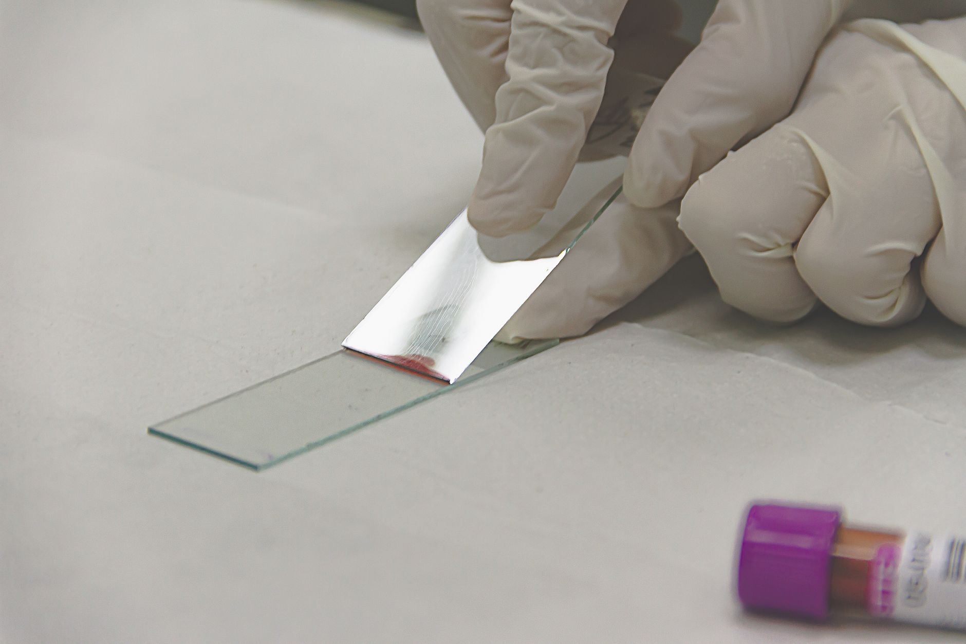
Somewhere in a freezer right now, a glass slide sits in a cardboard tray that nobody has touched in four years. The tissue on it is irreplaceable. The data on it is effectively invisible. That's the core problem digitization solves, and it's why biomedical research labs are rethinking how they store, share, and analyze physical samples at a fundamental level.
Glass slides have always been fragile, geography-dependent, and stubbornly analog. You hand-carry them to a microscope, one pathologist reviews one slide at a time, and collaboration across sites means shipping physical materials through a courier. For a field increasingly driven by multi-center studies and AI-powered analysis, that model just doesn't hold up.
What Whole Slide Imaging Actually Does to a Research Lab
Whole slide imaging, or WSI, converts a glass slide into a high-resolution digital file, typically called a whole slide image, that researchers can pan, zoom, annotate, and share the same way you'd work with any digital document. The difference is that each file can run to several gigabytes, capturing cellular detail at resolutions that rival what you'd see through an optical lens.
The practical payoff shows up fast in multi-site environments. WSI has emerged as a significant technology in clinical diagnostics, medical education, and pathology research, enabling advanced remote collaboration, the integration of AI into diagnostic workflows, and large-scale data sharing for multi-center research. That last point matters most for pharmaceutical and biotech research teams running coordinated studies across labs in different cities or countries.
Consider a concrete example. A contract research organization running a tumor morphology study across three sites in Boston, Amsterdam, and Singapore used to ship physical slides between teams, losing two to four days per review cycle and risking breakage in transit. After digitizing their glass archive, all three teams reviewed the same WSI files simultaneously through a shared annotation platform. Review cycles that once spanned a week compressed to a single business day. The slides never left the building.
The Efficiency Gap Research Teams Rarely Expect
There's a nuance that rarely makes it into vendor brochures. Early in the adoption curve, digital workflows can feel slower, not faster, because researchers are learning new tools and workflows at the same time. The gains come after full integration.
Research published on PubMed found that, taking into account case distribution, a digital workflow produced an average gain of 12.3% in reading time, with WSI instead of conventional microscopy significantly reducing review times overall. That 12.3% is a conservative average that masks much larger gains in specific categories. The same study recorded a 68% time reduction for prostate biopsies specifically, once the workflow was fully integrated.
The takeaway for lab managers: the efficiency argument for digitization is real, but it requires genuine system integration, not just buying a scanner and dropping files onto a shared drive. Your image management platform, your annotation tools, and your downstream analysis pipelines all need to talk to each other before the gains kick in.
AI Model Training: The Use Case Nobody Budgeted For
Three years ago, most biomedical labs digitized slides to solve a storage or collaboration problem. Today, AI model training has become an equally compelling reason, and in some research environments it's the primary one.
Deep learning models for tissue classification, biomarker detection, and tumor morphology analysis all require large, cleanly labeled image datasets. Glass slides don't feed those pipelines. High-resolution WSIs do, and the scale of training data needed makes digitization not optional but foundational. Labs that built digital archives early for mundane archival reasons are now sitting on datasets other teams are scrambling to assemble from scratch.
The adoption gap, though, is real. According to a 2024 estimate from the College of American Pathologists, digital pathology adoption sits at approximately 10% in U.S. labs. That figure likely reflects routine clinical diagnostics more than research environments, where adoption runs higher, but the number illustrates how much runway remains before WSI becomes the default. Research teams that move now are building institutional advantages that compound over time.
On the AI familiarity side, the picture is similarly split. A 2025 global survey published in The Journal of Pathology found that while 73% of respondents reported some familiarity with AI, actual use was limited, with 31% reporting rare use and 29% no use at all. Familiarity and active deployment are very different things, and the gap between them is largely a data infrastructure problem. You can't build AI workflows without digital images to train on.
For research teams that need pre-scanned tissue datasets or need their own glass slides converted into analysis-ready WSIs, working with established digital pathology services lets them skip the capital cost of scanner hardware and the operational overhead of building an in-house imaging lab.
The Digitization Decision Matrix
Not every lab digitizes for the same reason. Before committing budget, it helps to map your primary driver against the infrastructure investment each use case actually demands. Here's a framework, the Digitization Decision Matrix, that clarifies that trade-off:
|
Primary Use Case |
WSI Resolution Needed |
Storage Burden |
Best Acquisition Path |
|
Remote collaboration and review |
20x (standard) |
Moderate (500MB to 1GB per slide) |
External scanning service |
|
AI and deep learning model training |
40x (high) |
High (1GB to 3GB per slide) |
External scanning service with format flexibility |
|
Rare case archive preservation |
20x to 40x depending on tissue type |
Moderate to high |
External scanning service (one-time batch) |
|
High-volume routine research |
20x to 40x |
Very high |
In-house scanner plus cloud storage infrastructure |
The matrix reveals something counterintuitive: the most expensive-feeling path, buying an in-house scanner, is only justified when volume is consistently high and turnaround time is a daily bottleneck. For most academic and biotech research teams running project-based studies, external scanning covers the need at a fraction of the capital outlay.
File Formats, Software Compatibility, and the Portability Problem
One underappreciated friction point is file format lock-in. Different scanners produce proprietary formats, and not every annotation or analysis platform can read every format natively. SVS, NDPI, SCN, and MRXS are common, but cross-platform workflows often require conversion steps that add time and occasionally degrade metadata.
The practical fix is to specify format requirements before you commission any scanning work. If your downstream pipeline runs on a specific viewer or AI platform, confirm format compatibility upfront. Most commercial scanning providers can deliver in multiple formats, but only if you ask before the work starts, not after you receive 400 files you can't open.
What Research Teams Get Wrong About Archive Conversion
Archive conversion projects, converting years of stored glass slides into digital files, routinely underestimate two costs: quality control and metadata management. A WSI file without accurate slide metadata is almost useless for research purposes. Tissue type, stain protocol, patient cohort identifiers, and case numbers all need to travel with the image file in a structured, searchable format.
The labs that run archive projects successfully treat metadata schema design as a deliverable equal in importance to the scan itself. They define their data dictionary first, then scan. Teams that scan first and figure out metadata later end up with digital files that are just as difficult to work with as the physical slides they replaced.
The Road Ahead for Research Labs
Glass slides aren't disappearing anytime soon. The physical tissue on a glass slide remains the irreplaceable source of truth. But the workflow built around that slide, the manual microscopy review, the physical shipping, the one-at-a-time analysis, is genuinely being replaced by faster, more scalable digital alternatives.
Research teams that treat digitization as a one-time IT project tend to underinvest in it. The labs seeing the strongest return treat it as an ongoing data strategy: building archives that grow more valuable as AI tools improve, maintaining format flexibility as platforms evolve, and building institutional knowledge about how to manage image data at scale.
The microscope isn't obsolete. But the workflow around it? That's already changed. The question isn't whether your lab will make the shift, it's whether you'll do it before your collaborators do it first.